Your dentist might spot something unusual in your mouth during a routine exam, like a white patch, red area, or sore that won’t heal. When this happens, a biopsy becomes one of the most important tools for figuring out what’s going on.
A dental biopsy provides an accurate diagnosis by examining tissue samples, which helps detect serious conditions like oral cancerin their earliest and most treatable stages.
The process might sound scary, but understanding how biopsies help detect oral disease can give you peace of mind. Most biopsies are quick procedures done right in your dentist’s office with local anesthesia. The tissue sample gets sent to a lab where experts look at it under a microscope to identify any problems.
Early detection through oral biopsies can make a huge difference in your treatment options and overall health outcomes. Many oral lesions turn out to be harmless, but getting a definitive answer helps you avoid worry and catch any serious issues before they progress.
Key Takeaways
- Biopsies provide accurate diagnoses of oral lesions by examining tissue samples under a microscope
- Early detection through biopsy improves treatment success and can identify serious conditions before they advance
- Most oral biopsy procedures are simple, low-risk, and performed quickly in a dental office with local anesthesia
The Importance of Early Detection in Oral Disease
Early detection of oral diseases can make the difference between simple treatment and complex interventions. Catching problems early improves your chances of successful treatment and helps identify if you’re at higher risk for serious conditions.
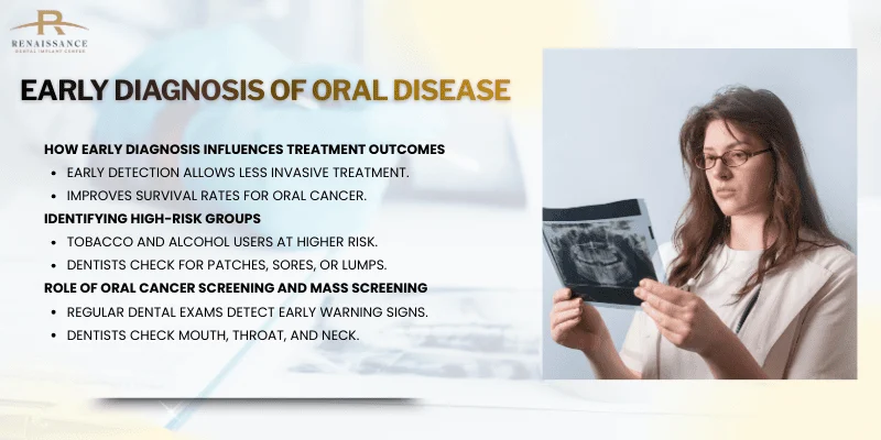
How Early Diagnosis Influences Treatment Outcomes
When your dentist finds oral diseases early, your treatment options are usually less invasive and more effective. Early oral cancer detection allows doctors to use simpler procedures with better survival rates. You might only need minor surgery instead of extensive treatments that affect your ability to eat or speak.
Early intervention significantly improves patient prognosis because doctors can treat the problem before it spreads. For oral cancer, finding it in stage one or two means you have a much better chance of recovery. Your treatment costs less, takes less time, and causes fewer side effects.
Early diagnosis also prevents complications that come with advanced disease. You avoid more painful procedures and longer recovery times. Your oral health stays better overall when problems get caught quickly.
Identifying High-Risk Groups for Oral Diseases
Some people face higher risks for oral diseases and need more careful monitoring. If you use tobacco products or drink alcohol regularly, your risk of oral cancer increases significantly. People over 40 should pay extra attention to changes in their mouth.
Your dentist looks for early signs during regular checkups. These include white or red patches, sores that don’t heal, or lumps in your mouth. If you have a family history of oral cancer or immune system problems, you need more frequent screenings.
Dental professionals are uniquely positioned to perform cancer screening exams during your routine visits. They can spot potentially dangerous lesions before you notice symptoms. Getting checked regularly is especially important if you fall into any high-risk category.
Role of Oral Cancer Screening and Mass Screening
Regular oral cancer screening during dental visits helps catch problems you might miss. Your dentist examines all areas of your mouth, throat, and neck for suspicious changes. This simple exam takes just a few minutes but can save your life.
Mass screening programs reach larger populations, especially in areas where people lack regular dental care. Brush biopsies are suitable as a non-invasive automated oral cancer screening tool for mass screening because they cost less and cause no pain. These programs help detect oral cancer in communities that need it most.
You should ask your dentist about oral cancer screening at every checkup. The exam looks for early warning signs that could indicate cancer or precancerous conditions. Catching these changes early gives you the best chance for successful treatment.
Understanding Oral Biopsy Procedures
An oral biopsy involves removing a small tissue sample from your mouth so a specialist can examine it under a microscope. The procedure uses local anesthesia to keep you comfortable, and knowing what to expect helps reduce anxiety about the process.
When Is a Biopsy Recommended?
Your dentist or oral surgeon may recommend a biopsy when they notice unusual changes in your mouth that need further investigation. Non-healing sores that last more than two weeks are a common reason for biopsy. These sores don’t respond to normal treatment and could indicate a serious condition.
White or red patches that appear on your gums, tongue, or cheeks often require examination. Lumps or bumps that develop in your oral tissues also need evaluation through biopsy procedures.
You might need a biopsy if you experience difficulty swallowing or persistent pain in your mouth without a clear cause. Changes in tissue color or texture that your dentist finds concerning warrant further testing. Early detection through biopsies can identify problems before they become more serious health issues.
Step-by-Step Overview of the Biopsy Procedure
The biopsy starts when your oral surgeon examines the area and decides which technique to use. They clean the site thoroughly before beginning any work.
Your doctor numbs the area completely before removing any tissue. The actual sample collection takes only a few minutes once you’re numb. For small growths, the surgeon may remove the entire lesion through an excisional biopsy. Larger areas require an incisional biopsy where only a portion is removed.
A brush biopsy offers a less invasive option for superficial tissue. Your surgeon uses a small brush to collect cells from the surface. After removing the tissue, they may place a few stitches to help the area heal properly.
The tissue sample goes to a laboratory where specialists examine it under microscopes. Results typically come back within a few days to a week.
Importance of Local Anesthesia and Patient Preparation
Local anesthesia keeps you comfortable during the entire biopsy procedure. Your oral surgeon injects the numbing medication into the tissue around the area being examined. You’ll feel pressure but no pain during the procedure.
Before your appointment, you may need to avoid eating or drinking for a few hours. Tell your oral surgeon about any medications you take or health conditions you have. Some medicines can affect bleeding or healing.
Follow any oral hygiene instructions your doctor gives you before the procedure. Clean teeth and gums help reduce infection risk. Most patients feel minimal discomfort because the anesthesia works effectively.
Post-Biopsy Care and Recovery
After your biopsy, you’ll need to follow specific care instructions to promote healing. Some minor bleeding or swelling is normal in the first day or two. Apply gentle pressure with clean gauze if bleeding occurs.
Avoid hot foods and drinks for the first 24 hours. Stick to soft foods that won’t irritate the biopsy site. Don’t brush directly over the area until your surgeon says it’s okay.
Take any prescribed pain medication as directed. Over-the-counter pain relievers usually handle any discomfort. Watch for signs of infection like increased swelling, severe pain, or fever. Contact your oral surgeon if these symptoms develop.
Most people recover quickly from oral biopsies and return to normal activities within a few days.
Types of Biopsy Techniques for Early Oral Disease Detection
Different biopsy methods allow your dentist to collect samples from suspicious oral lesions, ranging from removing entire small lesions to collecting cells with a brush. Tissue biopsy and liquid biopsy represent the two main categories, each offering distinct advantages for detecting early oral disease.
Incisional and Excisional Biopsy Explained
An incisional biopsy removes only a portion of a suspicious lesion for testing. Your doctor uses this technique when the lesion is too large to remove completely or when removing it entirely might cause complications.
An excisional biopsy removes the entire lesion along with a small margin of healthy tissue around it. This approach works best for small lesions under 1 centimeter. You get both diagnosis and treatment in one procedure if the lesion turns out to be benign.
Punch biopsy is a common type of incisional biopsy. Your dentist uses a circular blade to remove a small cylinder of tissue from the lesion. This method causes minimal bleeding and typically requires only a few stitches.
Surgical biopsy provides the most accurate assessment for localized lesions. However, these procedures are invasive and require greater technical effort compared to other sampling methods.
Brush Biopsy and Cytobrush
Brush biopsy uses a small, stiff brush to collect cells from the surface and deeper layers of oral lesions. You experience minimal discomfort during this procedure since it doesn’t require anesthesia or cutting.
The cytobrush technique specifically targets cells from all three layers of the oral epithelium. Your dentist rotates the brush against the lesion until pinpoint bleeding appears, indicating they’ve reached the deeper tissue layers.
This method works well as a screening tool for lesions that don’t look obviously cancerous. The collected cells are sent to a lab where specialists examine them under a microscope for abnormalities.
Brush biopsy is less invasive than surgical options and doesn’t cause significant bleeding. However, if suspicious cells are found, you’ll still need a traditional tissue biopsy for confirmation.
Fine Needle Aspiration and Liquid-Based Cytology
Fine needle aspiration uses a thin, hollow needle to extract cells or fluid from lumps or masses in your mouth or neck. Your doctor often uses this technique to sample lymph nodes that might contain cancer cells.
The procedure takes just a few minutes and usually doesn’t require anesthesia. Your doctor inserts the needle multiple times to collect adequate cells from different areas of the suspicious tissue.
Liquid biopsy involves analyzing body fluids like saliva, blood, or urine for cancer biomarkers. These samples are easily collected and transported, making them ideal for screening and monitoring disease progression.
Liquid-based cytology preserves collected cells in a special solution rather than smearing them directly onto slides. This technique reduces artifacts and allows for additional molecular testing on the same sample.
The main limitation is lower sensitivity compared to tissue biopsy. You’ll need an initial pathological diagnosis before liquid biopsy can effectively track your condition over time.
Emerging Biopsy Techniques and Technologies
Autofluorescence imaging helps your dentist identify lesion margins before taking a biopsy. Abnormal tissues appear darker under special blue light compared to healthy tissue, which glows green.
Next-generation sequencing (NGS) analyzes multiple genes simultaneously from biopsy samples. This technology detects genetic mutations associated with oral cancer, even in very small tissue samples.
Near-infrared imaging with special dyes allows surgeons to visualize lymph nodes and tumor margins more clearly during procedures. Better visualization leads to more accurate sample collection from suspicious areas.
Molecular-specific fluorescent contrast agents and spectroscopy help identify cancerous changes at the cellular level. These techniques may soon allow real-time assessment during biopsy procedures, reducing the need for multiple visits.
Electrochemical assays detect specific cancer markers like telomerase activity in biopsy samples. These rapid tests provide results faster than traditional methods, though they’re still being refined for clinical use.
Analyzing and Interpreting Biopsy Results
After your biopsy sample is collected, pathologists examine the tissue using specialized techniques to identify disease markers and abnormalities. The accuracy of these findings depends on advanced methods like histopathological examination and molecular testing that can detect early signs of conditions like oral squamous cell carcinoma.
Role of the Pathologist
Your pathologist specializes in examining tissue samples removed during biopsies. They look at cells under a microscope to identify disease patterns and abnormalities that indicate specific conditions.
Surgical pathologists focus on tissues from biopsies, while cytopathologists analyze individual cells from procedures like fine needle aspiration. These specialists have training in recognizing subtle changes in cell structure and tissue organization.
Your pathologist writes a detailed report that includes your patient information, the type of specimen collected, and specific findings from the analysis. This report guides your doctor in making treatment decisions and planning follow-up care.
The pathologist’s interpretation forms the foundation for your diagnosis and helps your healthcare team understand whether disease is present and how aggressive it might be.
Histopathological Examination and Histopathology
Histopathological examination involves analyzing your tissue sample under a microscope to look for abnormal cell patterns and structural changes. Your tissue goes through processing steps that include preservation, slicing into thin sections, and staining with special dyes.
These stains highlight different cell components and make it easier to see abnormalities. The pathologist examines the tissue architecture, cell size and shape, and how cells relate to surrounding structures.
For oral disease detection, histopathology can reveal precancerous changes called dysplasia or confirm the presence of malignant cells. The examination shows specific features like:
- Cell irregularity and abnormal growth patterns
- Changes in tissue layers and boundaries
- Presence of inflammatory cells or infection
- Degree of cellular differentiation
Your histopathology results often include a grade or stage that indicates disease severity.
Use of Biomarkers and Molecular Diagnostics
Biomarkers are specific molecules in your cells that indicate disease presence or progression. Modern biopsy analysis includes molecular testing that looks beyond what’s visible under a microscope.
NGS (next-generation sequencing) technology can identify genetic mutations and molecular changes in your tissue sample. This advanced testing provides information about specific genetic alterations that drive disease development.
For oral squamous cell carcinoma, molecular diagnostics can detect mutations in genes like TP53 and identify proteins that indicate cancer risk. These tests help predict how your disease might behave and which treatments will work best.
Molecular assessment techniques improve the sensitivity and accuracy of biopsy analysis compared to traditional methods alone. Your doctor uses this molecular information alongside standard histopathology to create a complete picture of your condition.
Diagnostic Accuracy and Clinical Utility
The diagnostic accuracy of your biopsy depends on proper sample collection, handling, and analysis techniques. When performed correctly, biopsies provide reliable information for diagnosing oral diseases and planning treatment.
Biopsy results play a crucial role in determining your treatment path, whether that means starting therapy, monitoring disease progression, or confirming absence of disease. Your healthcare team relies on these findings to make evidence-based decisions.
The clinical utility extends beyond initial diagnosis. Repeat biopsies help track how well your treatment is working and detect any disease recurrence early.
Accuracy improves when pathologists use standardized protocols and combine traditional histopathology with molecular testing. Your biopsy results give specific information about disease type, extent, and biological behavior that imaging tests alone cannot provide.
Biopsies in Identifying Oral Lesions and Potentially Malignant Disorders
Biopsies help dentists identify which oral lesions need immediate treatment and which ones might turn into cancer over time. Early identification of oral cancer development leads to better treatment outcomes and can prevent changes from benign to malignant.
Recognizing Leukoplakia, Erythroplakia, and Mucosal Lesions
Leukoplakia appears as white patches in your mouth that won’t scrape off. These patches show up most often on your tongue, inside your cheeks, or on your gums. Leukoplakia has a global prevalence of 1.7-2.7% with a malignant conversion rate of approximately 1.36%.
Erythroplakia looks like red patches or spots in your mouth. This condition is less common than leukoplakia but carries a higher risk of becoming cancer.
Your dentist will recommend a biopsy if these mucosal lesions last longer than two weeks. Oral biopsies are recommended for lesions that persist after potential irritants are removed. The biopsy removes a small piece of tissue so a specialist can examine it under a microscope.
Oral Epithelial Dysplasia and Lichen Planus
Oral epithelial dysplasia means the cells in your mouth tissue look abnormal under a microscope. Your doctor grades this condition as mild, moderate, or severe based on how different the cells appear from normal tissue. Severe dysplasia has a higher chance of turning into cancer.
Oral lichen planus creates white, lacy patterns or red, swollen tissues in your mouth. This chronic condition can cause discomfort and rarely develops into cancer. Dental hygienists play a crucial role in detecting suspicious lesions, whether they’re benign, precancerous, or cancerous.
Your dentist uses tissue biopsies to confirm these conditions. The biopsy results guide your treatment plan and help determine how often you need follow-up appointments.
Diagnosis of Oral Potentially Malignant Disorders
Oral potentially malignant disorders include several conditions that might develop into cancer. These include leukoplakia, erythroplakia, oral lichen planus, oral submucous fibrosis, and proliferative verrucous leukoplakia.
Meta-analysis shows that 7.9% of oral potentially malignant disorder cases turn into carcinoma with moderate or severe dysplasia. Your dentist performs a biopsy to assess your risk level and create a monitoring schedule.
The biopsy tells your doctor if the tissue shows dysplasia and how severe it is. This information helps predict which lesions need aggressive treatment versus careful watching. Clinical guidelines recommend that clinicians perform a biopsy or provide immediate referral to a specialist for suspicious oral mucosal lesions.
Monitoring Persistent Oral Symptoms
You should see your dentist if you notice sores, lumps, or color changes in your mouth that don’t heal within two weeks. Persistent symptoms include patches that bleed easily, areas that feel hard or fixed to deeper tissues, or lesions that grow rapidly.
Your dentist examines your mouth during routine checkups and looks for changes in existing lesions. Regular monitoring helps catch problems early when treatment works best.
Warning signs that need a biopsy:
- Red or mixed red and white patches
- Ulcers that don’t heal
- Lumps or thick areas
- Areas that bleed without cause
- Numbness or pain that persists
Most early-stage oral cancer cases are asymptomatic, which makes regular dental visits critical. Your dentist might use special lights or dyes to spot abnormal tissue before performing a biopsy.
Future Perspectives in Oral Disease Detection
New technologies are transforming how doctors find oral diseases early, with methods that are less painful and more accurate. Liquid biopsy and advanced imaging can detect problems before symptoms appear, while artificial intelligence helps identify diseases faster than traditional approaches.
Non-Invasive and Minimally Invasive Biopsy Methods
You no longer need to worry as much about painful procedures when checking for oral disease. Scientists are developing biopsy techniques that don’t require cutting into your tissue or cause discomfort.
Brush biopsies let your dentist collect cells by gently brushing the suspicious area in your mouth. This approach works well for initial screening before deciding if you need a traditional biopsy.
Optical coherence tomography creates detailed images of your oral tissues without touching them. The technology uses light waves to see below the surface, helping doctors spot abnormal cells early.
Liquid biopsy methods analyze your blood or saliva instead of tissue samples. These tests look for circulating tumor DNA (ctDNA) that cancer cells release into your body fluids.
Advances in Autofluorescence and Imaging
Autofluorescence devices shine special blue light into your mouth to make abnormal tissues glow differently than healthy ones. When your dentist uses this tool, cancerous or precancerous areas appear darker because they don’t reflect light the same way normal tissue does.
Key imaging improvements include:
- Digital imaging systems that capture clearer pictures of suspicious areas
- Spectroscopy tools that analyze how light interacts with your tissue
- Narrow-band imaging that highlights blood vessel patterns in abnormal growths
These technologies help your dentist see problems that the naked eye might miss during regular exams. Visual inspection alone often fails to detect oral cancers early, making these advanced tools valuable for catching disease sooner.
Role of Salivary Biomarkers and Liquid Biopsy
Your saliva contains biomarkers that reveal early signs of oral disease. Scientists can test your spit for specific proteins, DNA fragments, and RNA molecules that indicate cancer or other problems.
High-throughput sequencing and digital PCR have made these tests more sensitive and accurate. The technology detects tiny amounts of abnormal genetic material before you develop symptoms.
Liquid biopsy tracks ctDNA levels to monitor how well your treatment is working. If levels drop, your therapy is effective. Rising levels may signal that the disease is coming back.
Benefits of salivary testing:
- No needles or tissue removal needed
- Easy to repeat for ongoing monitoring
- Results available faster than traditional biopsies
Integration of Artificial Intelligence and Digital Pathology
Artificial intelligence systems can detect oral cancer by analyzing images of your mouth with impressive accuracy. These programs learn from thousands of cases to spot patterns that humans might overlook.
Next-generation sequencing (NGS) combined with AI helps doctors understand the genetic makeup of your oral disease. This information guides treatment decisions based on your specific type of cancer.
Digital pathology lets multiple experts review your biopsy slides electronically. AI software can highlight suspicious cells and suggest diagnoses, speeding up the process while reducing errors.
Machine learning algorithms also help predict which oral lesions will turn cancerous. Your dentist can use this information to decide how aggressively to monitor or treat your condition.
Frequently Asked Questions
Oral biopsies help identify many conditions beyond cancer, and the procedure itself involves several straightforward steps. Understanding the different types of biopsies and what to expect can help ease concerns about this important diagnostic tool.
What conditions can a mouth biopsy help diagnose?
A mouth biopsy can identify a wide range of oral health problems. Biopsies help diagnose benign conditions, infections, and abnormalities in oral tissues, not just cancer.
Your dentist or oral surgeon might recommend a biopsy to check for oral cancer or precancerous lesions. These procedures also detect inflammatory conditions and various infections that affect your mouth.
White patches called leukoplakia and red patches known as erythroplakia are common findings that need biopsy confirmation. Biopsies can also identify benign tumors like fibromas and help determine the cause of persistent ulcers or unusual growths in your mouth.
What is the most commonly performed biopsy in the oral cavity?
The incisional biopsy is one of the most frequently performed types in oral surgery. This method removes a small portion of suspicious tissue when the lesion is too large to take out completely.
Punch biopsies are also widely used because they work well for many situations. This technique uses a circular blade to remove a small cylinder of tissue, making it ideal for lesions that might involve deeper tissue layers.
The choice between biopsy types depends on the size and location of the abnormal area. Your oral surgeon will select the method that provides the best tissue sample for accurate diagnosis.
Can you walk me through the oral biopsy procedure?
Your oral surgeon starts with a thorough examination of your mouth to identify the suspicious area. Before removing any tissue, the area gets numbed with local anesthesia to keep you comfortable during the procedure.
Once you’re numb, your surgeon uses the chosen biopsy technique to collect the tissue sample. The sample is then sent to a pathology laboratory where a specialist examines it under a microscope.
After the biopsy, you’ll receive instructions for taking care of the area. These typically include guidance on pain management and keeping your mouth clean while it heals.
Which techniques are commonly used for conducting an oral biopsy?
Four main biopsy techniques are used to examine oral tissue. Each method serves a specific purpose based on the characteristics of the lesion.
Incisional biopsies remove part of a larger lesion for testing. Excisional biopsies take out the entire suspicious area, serving both diagnostic and treatment purposes.
Punch biopsies use a circular tool to extract a small cylinder of tissue. Brush biopsies collect cells from the surface using a small brush, though this less invasive method may not always provide enough tissue for a complete diagnosis.
How many different types of oral biopsies are there?
There are four main types of oral biopsies used in dental practice. Incisional, excisional, punch, and brush biopsies each offer different advantages depending on your specific situation.
Incisional and excisional biopsies are the traditional surgical approaches. Punch biopsies provide a middle ground for accessing deeper tissue layers.
Brush biopsies represent the least invasive option. Advanced techniques like laser biopsies are also becoming more common, offering precise tissue removal with less bleeding and discomfort.
What is the usual timeframe for receiving mouth biopsy results?
Most biopsy results come back within one to two weeks after your procedure. The exact timing depends on the complexity of the analysis and the workload at the pathology laboratory.
Your oral surgeon will schedule a follow-up appointment to discuss the results with you. During this visit, they’ll explain what the biopsy found and recommend next steps if treatment is needed.
Some cases require additional testing or specialized analysis, which can extend the waiting period. Your dental team will keep you informed if any delays occur in processing your biopsy results.